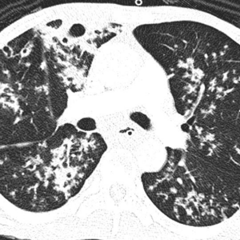
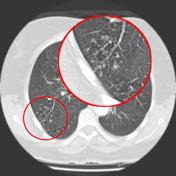
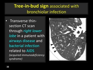
tree in bud opacities seen in
They are suggestive for the diagnosis of congestive heart failure but are also seen in various non-cardiac conditions such as pulmonary fibrosis interstitial deposition of heavy metal particles or carcinomatosis of the lung. Bronchiolar dilatation often referred to as bronchiolectasis mosaic attenuation andor air trapping if expiratory imaging is used Classification.
The acute course of COVID-19 is variable and ranges from asymptomatic infection to fulminant respiratory failure.

. Adjacent bronchial wall thickening is also frequently depicted. Associated focal ground-glass and consolidative opacities may be visualized although this should not the predominant feature. Classically bronchiolitis appears as a region of centrilobular nodularity often in a tree-in-bud pattern.
MAC has been reported as the most common organism in these patients and is responsible for an indolent infectious process 2 7 8. Chest CT showed a filling defect in the left atrium and a mobile left atrial mass was seen on transthoracic echocardiography shown in a. Multiple causes for tree-in-bud TIB opacities have been reported.
It is a non-specific sign with a wide etiology including infection chronic interstitial disease and acute alveolar disease. Most common radiological findings seen on chest CT include scattered centrilobular nodules tree-in-bud opacities and fibronodular bronchiectasis. However to our knowledge the relative frequencies of the causes have not been evaluated.
It is usually visible on standard CT however it is best seen on HRCT chest. Patients recovering from COVID-19 can have persistent symptoms and CT abnormalities of variable severity. A 47-year-old woman presented with shortness of breath.
Tree-in-bud sign is not generally visible on plain radiographs 2. In the right mid-lung nodular opacities are in a tree-in-bud distribution suggestive of endobronchial spread. Coronal reconstructed computed tomography image shows the lingular cavity with irregular nodules and right mid-lung nodular opacities in a 43-year-old man who.
In the absence of primary lung pathology. Typically the centrilobular nodules are 2-4 mm in diameter and peripheral within 5 mm of the pleural surface. TIB opacities were seen in association with bronchiectasis in 30 123 of 406 of cases and with an apical-predominant disease in 25 10 of 406 cases.
Ground-glass opacificationopacity GGO is a descriptive term referring to an area of increased attenuation in the lung on computed tomography CT with preserved bronchial and vascular markings. One method of classifying various forms of bronchiolitis is as follows 1. Centrilobular micronodules often seen as tree-in-bud opacities bronchial wall thickening.
Small patchy peripheral opacities are also present in the left lower lobe. In the remaining 67 273 of. Chronic Kerley B lines may be caused by fibrosis or hemosiderin deposition caused by recurrent pulmonary edema.
High-resolution CT scan of the thorax demonstrates central bronchiectasis a hallmark of allergic bronchopulmonary aspergillosis right arrow and the peripheral tree-in-bud appearance of centrilobular opacities left arrow which represent mucoid impaction of the small bronchioles. At 3 months after acute infection a subset of patients will have CT abnormalities that include ground-glass opacity GGO and subpleural bands with.
View Of Tree In Bud The Southwest Respiratory And Critical Care Chronicles

Ct Scan Of Chest Revealing Scattered Tree In Bud Opacities In Both Download Scientific Diagram

Tree In Bud Pattern Pulmonary Tb Eurorad

Tree In Bud Sign Lung Radiology Reference Article Radiopaedia Org

Tree In Bud Caused By Haemophilus Influenzae Radiology Case Radiopaedia Org

Tree In Bud Sign Golden S Sign

Tree In Bud Pattern Pulmonary Tb Eurorad
View Of Tree In Bud The Southwest Respiratory And Critical Care Chronicles

Tree In Bud Caused By Haemophilus Influenzae Radiology Case Radiopaedia Org

Tree In Bud Pattern Pulmonary Tb Eurorad

Pdf Tree In Bud Semantic Scholar

Hrct Scan Of The Chest Showing Diffuse Micronodules And Tree In Bud Download Scientific Diagram

Tree In Bud Sign Lung Radiology Reference Article Radiopaedia Org
